231


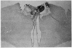
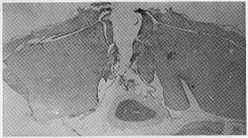
Fig. 1. On the left side of this figure brain damage sustained by the Al (ΛNT) and Pl
(POST) groups of Guinea fowl are reconstructed on five evenly spaced serial sections
from anterior (upper) to posterior (lower). This covers approximately 2 mm. Damage
sustained by all members of the group is shaded. The maximum degree of damage sustain-
ed by any bird at the given plane is represented by the hatched area. The figure ,n1 states
the number of birds with damage at this plane. On the right-hand side of the figure, the
two photographs show transverse sections in the plane of maximum damage for one ex-
ample from the Al (upper) and the Pl group (lower). For further details see text (x 4.13).
Aa: Archistriatum, pars anterior; Ai: Archistriatum, pars intermedium; APH: Area para-
hippocampalis; CA: Anteriorcommissure; FA: IVactus fronto-archistriatalis; HA: Hyper-
striatum accessorium; HD: Hyperstriatum dorsale; Hp: Hippocampus; HV: Hyperstriatum
ventrale; L: Field L; N: Neostriatum; PA: Paleostriatum augmentatum; PP: Paleostriatum
primitivum; S; Septum.
More intriguing information
1. An institutional analysis of sasi laut in Maluku, Indonesia2. A Pure Test for the Elasticity of Yield Spreads
3. Deprivation Analysis in Declining Inner City Residential Areas: A Case Study From Izmir, Turkey.
4. Computing optimal sampling designs for two-stage studies
5. Labour Market Flexibility and Regional Unemployment Rate Dynamics: Spain (1980-1995)
6. On the Relation between Robust and Bayesian Decision Making
7. Natural Resources: Curse or Blessing?
8. Does Competition Increase Economic Efficiency in Swedish County Councils?
9. The name is absent
10. The name is absent