Sponsored Document Sponsored Document Sponsored Document
Creese and Cooper
Page 18

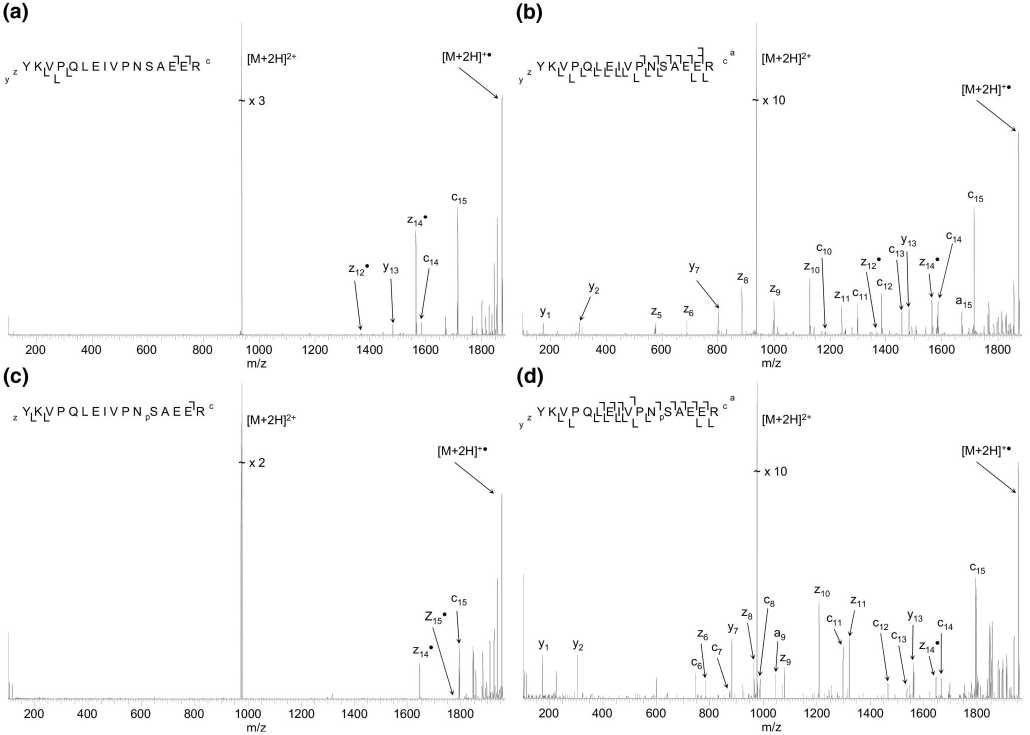
Figure 6.
(Top) ECD mass spectra (3 scans) of doubly-charged ions of the α-S1-casein tryptic peptide
YKVPQLEIVPNSAEER obtained at ECD cathode potentials (a) -3.34 V (standard) and (b)
-11.84 V. (Bottom) ECD mass spectra (5 scans) of doubly-charged ions of
YKVPQLEIVPNpSAEER obtained at cathode potentials of (c) -3.34 V (standard) and (d)
-12.34 V.
Published as: JAm SocMass Spectrom. 2008 September ; 19(9): 1263-1274.
More intriguing information
1. The name is absent2. CONSUMER PERCEPTION ON ALTERNATIVE POULTRY
3. The name is absent
4. THE CO-EVOLUTION OF MATTER AND CONSCIOUSNESS1
5. Aktive Klienten - Aktive Politik? (Wie) Läßt sich dauerhafte Unabhängigkeit von Sozialhilfe erreichen? Ein Literaturbericht
6. EXECUTIVE SUMMARY
7. Palvelujen vienti ja kansainvälistyminen
8. Effects of red light and loud noise on the rate at which monkeys sample the sensory environment
9. Evidence-Based Professional Development of Science Teachers in Two Countries
10. The name is absent