Sponsored Document Sponsored Document Sponsored Document
Veerapen et al.
Page 8

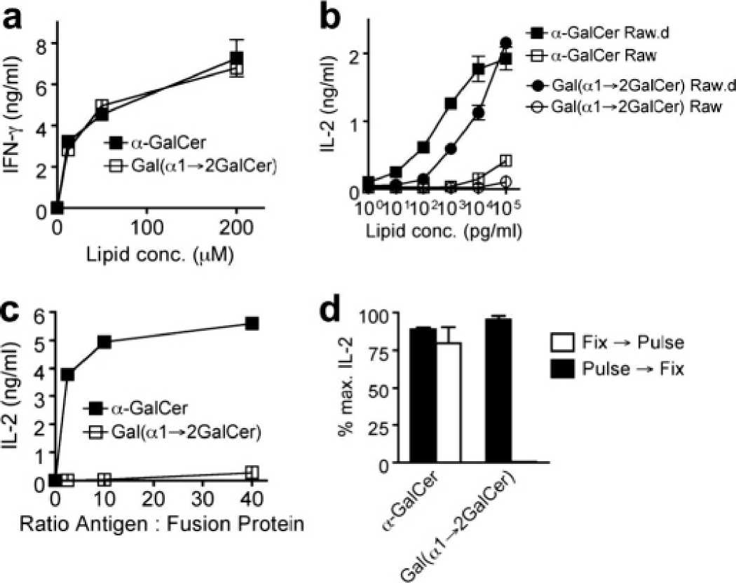
Figure 1.
α-GalCer and Gal(α1→2GalCer) stimulate CD1d-restricted iNKT cells. (a) In vitro activation
of 2.5 × 10⁴ human iNKT cells (clone BM2a.3) in co-culture with 2.5 × 10⁴ U937 cells and
various concentrations of α-GalCer (filled squares) and Gal(α1→2GalCer) (open squares).
After 16 h, cytokines were determined in culture supernatants by ELISA; (b) in vitro activation
of 5 × 10⁴ mouse iNKT cells (hybridoma DN32) in co-culture with 5 × 10⁴ RAW cells
transfected with CD1d (filled symbols) or untransfected (open symbols) and various
concentrations of α-GalCer (squares) and Gal(α1→2GalCer) (circles). After 16 h, cytokine
concentrations were determined in culture supernatants by ELISA; (c) Plate-bound murine
recombinant CD1d-Fc fusion proteins were loaded with α-GalCer (filled squares) or Gal
(α1→2GalCer) (open squares) for 16 h, washed, and 5 × 10⁴iNKT cell hybridomas were added
per well. Cytokines were determined in culture supernatants by ELISA; (d) RAW cells
transfected with CD1d were pulsed with 100 ng/ml of α-GalCer or Gal(α1→2GalCer) for 3 h
and then washed and fixed with glutaraldehyde (filled bars), or fixed and then pulsed for 3 h
(open bars). 105 APCs were co-cultured with 105iNKT cell hybridomas for 16 h and cytokines
were determined in culture supernatants by ELISA. Cytokine responses are expressed as
percent of maximal response. Methods are described elsewhere.
Published as: BioorgMed Chem Lett. 2009 August 01; 19(15): 4288-4291.
More intriguing information
1. The name is absent2. Feeling Good about Giving: The Benefits (and Costs) of Self-Interested Charitable Behavior
3. The name is absent
4. Valuing Access to our Public Lands: A Unique Public Good Pricing Experiment
5. Reversal of Fortune: Macroeconomic Policy, International Finance, and Banking in Japan
6. The name is absent
7. The name is absent
8. Philosophical Perspectives on Trustworthiness and Open-mindedness as Professional Virtues for the Practice of Nursing: Implications for he Moral Education of Nurses
9. CREDIT SCORING, LOAN PRICING, AND FARM BUSINESS PERFORMANCE
10. Putting Globalization and Concentration in the Agri-food Sector into Context