61
the posterior superior temporal sulcus where it angles upwards towards the parietal
lobe. The anatomical positioning of MT, MST and STSms in human cortex was similar to
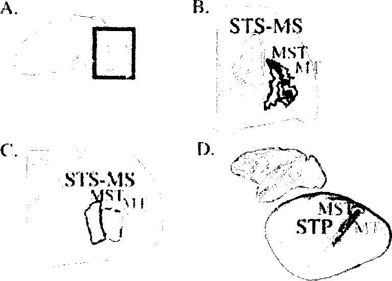
that of MT, MST and STP in macaque cortex (Fig. 5D).

Figure 5. Relationship between the STS multisensory area (STSms) and areas MT and MST. A. Lateral
view of a single subject's partially inflated left hemisphere. Colored regions responded significantly
to all three modalities. Active regions in posterior STS are colored yellow, other active regions are
colored purple. The fundus of the STS is shown as a white dashed line. Red box indicates the region
enlarged in B. B. Composite map showing multisensory activation and Iocalizer defined MT and
MST. White outline shows STSms, blue outline shows MST, green outline shows MT. C. Composite
map in an additional hemisphere from a different subject. D. Relationship between macaque area
STP and macaque areas MTand MST. The top panel shows a lateral view of a macaque brain
(Dickson, et al. 2001). The fundus of the STS is shown as a white dashed line. The bottom panel
shows an inflated view of the brain, with labeled areas from (Lewis and Van Essen 2000b): MT, MST
(MSTdp+MSTm) and STP (TPOi+TPOc).
Discussion
Guided by the literature on macaque STP, we hypothesized that human STS
should contain an area that responds to somatosensory, auditory and visual stimulation.
More intriguing information
1. The name is absent2. Monetary Discretion, Pricing Complementarity and Dynamic Multiple Equilibria
3. The name is absent
4. Credit Market Competition and Capital Regulation
5. Developments and Development Directions of Electronic Trade Platforms in US and European Agri-Food Markets: Impact on Sector Organization
6. he Effect of Phosphorylation on the Electron Capture Dissociation of Peptide Ions
7. Computational Batik Motif Generation Innovation of Traditi onal Heritage by Fracta l Computation
8. The name is absent
9. The name is absent
10. Bridging Micro- and Macro-Analyses of the EU Sugar Program: Methods and Insights