The deviations in the tip position during scanning are measured and correlated with
surface topography and a 3D image of the surface is reconstructed. The most commonly
used AFM systems rely on a laser bounced off the AFM tip assembly onto a spilt
photodiode to detect the variations in tip position (Figure 1.1), the commercial AFM used
in our studies also employed this system. As the tip is scanned across the sample surface,
the tip undergoes deformations which result in the laser beam spot being deflected. The
split photodiode can detect the beam spot movement by differential signals from its
different sections.
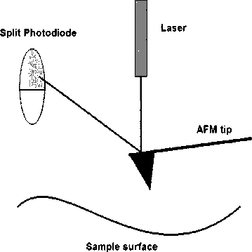
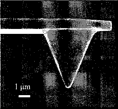
Figure 1.6: (Left) Schematic of an AFM optical detection system; a laser is bounced off
the back of the AFM tip and directed to a split photodiode. (Right) Scanning Electron
Microscope image of an AFM tip.
AFM is able to image in fluid and as a result, is able to image biological structures under
near-native conditions. AFM has been used to image cells [11], DNA strands [12],
antibodies [13] and many other biological structures.
More intriguing information
1. Linking Indigenous Social Capital to a Global Economy2. FDI Implications of Recent European Court of Justice Decision on Corporation Tax Matters
3. sycnoιogιcaι spaces
4. What should educational research do, and how should it do it? A response to “Will a clinical approach make educational research more relevant to practice” by Jacquelien Bulterman-Bos
5. Rent Dissipation in Chartered Recreational Fishing: Inside the Black Box
6. The name is absent
7. A Study of Adult 'Non-Singers' In Newfoundland
8. Howard Gardner : the myth of Multiple Intelligences
9. Do imputed education histories provide satisfactory results in fertility analysis in the Western German context?
10. Whatever happened to competition in space agency procurement? The case of NASA