Gelphase p., .j l
Fluid phase
≡≡β≡≡ ≡≡≡
Figure 3.13: Schematic of gel phase (blue) and fluid phase (yellow) lipid bilayers
coexisting. The gel phase lipid regions are more rigidly packed and are thicker than the
loosely packed fluid phase lipid regions
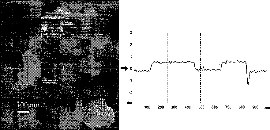
Figure 3.14: AFM imaging data of the DOPC:DOPS:DMPC:DMPS mixture. (Left)
Topography of the lipid mix, the lighter colors represent taller features - the image is Ixl
μm in size. (Right) A cross section taken through the topographical image - as shown by
the white line. The height difference between the lighter and the darker areas is 0.8 run,
identifying the lighter regions as gel phase patches residing in a sea of fluid phase lipid
sea.
In Figure 3.14, the topographical data clearly shows lighter areas which are 0.8 nm taller
than the surrounding lipid. These are identifiable as the gel phase lipid patches of
DMPC:DMPS located in a sea of fluid phase DOPC:DOPS. In order to compare the lipid
mobility based charge regulation of the gel and fluid lipids, we needed to have the surface
43
More intriguing information
1. Internationalization of Universities as Internationalization of Bildung2. An Interview with Thomas J. Sargent
3. Correlation Analysis of Financial Contagion: What One Should Know Before Running a Test
4. The name is absent
5. The name is absent
6. Human Resource Management Practices and Wage Dispersion in U.S. Establishments
7. Stakeholder Activism, Managerial Entrenchment, and the Congruence of Interests between Shareholders and Stakeholders
8. PER UNIT COSTS TO OWN AND OPERATE FARM MACHINERY
9. Dementia Care Mapping and Patient-Centred Care in Australian residential homes: An economic evaluation of the CARE Study, CHERE Working Paper 2008/4
10. PRIORITIES IN THE CHANGING WORLD OF AGRICULTURE