33
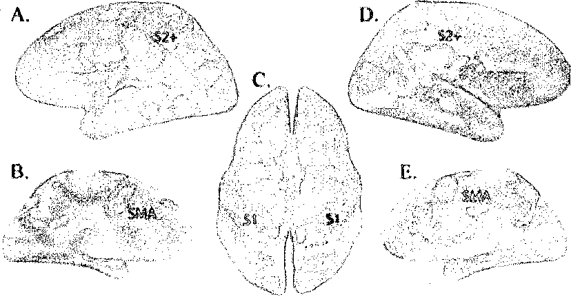
together discrete activation foci (S2+, STS, MST, and LOC) into a single large patch of
activation. The second largest area of activation was observed dorsally, in primary
somatosensory cortex (Sl) and adjacent areas, especially in a region in the postcentral
sulcus. On the medial wall of the hemisphere, tactile responses were found in the
supplementary motor area (Lim, et al. 1994) and posteriorly in the medial portion of SI.

Figure 5. Group map of cortical tactile activations with spatial smoothing (8mmfull-width half-
maximum Gaussian kernel). A, Lateral view of left hemisphere. B, Medial view of left hemisphere. C,
Superior view of both hemispheres. D, Lateral view of right hemisphere. E, Medial view of right
hemisphere. Outline shows selected regions. SMA1 Supplementary motor area, located in the medial
superior frontal cortex. Light green dashed line shows the fundus of the central sulcus, and dark
green dashed line shows the fundus of the postcentral sulcus.
Table 1. Clustered-node analysis of the group average surface activation map shown in Figure 5
|
Area |
Mean t |
Maximum t |
Coordinates c |
>f Location of | |
|
Left hemisphere Lateral occipital-temporal-parietal |
4545 |
4.9 |
18.2 |
(-54,-49,14) |
Posterior superior temporal sulcus |
|
Postcentral gyrus |
1834.25 |
4.4 |
8.7 |
(-15,-51,-59) |
Postcentral gyrus and sulcus |
|
Right hemisphere Lateral occipital-temporal-parietal |
2908 |
4.8 |
12.7 |
(40,-69,-2) |
Lateral occipital (MST) |
|
Postcentral gyrus |
1588 |
4.7 |
17.5 |
(21,-52,65) |
Postcentral gyrus |
More intriguing information
1. A Principal Components Approach to Cross-Section Dependence in Panels2. Keystone sector methodology:network analysis comparative study
3. Philosophical Perspectives on Trustworthiness and Open-mindedness as Professional Virtues for the Practice of Nursing: Implications for he Moral Education of Nurses
4. The name is absent
5. The name is absent
6. LAND-USE EVALUATION OF KOCAELI UNIVERSITY MAIN CAMPUS AREA
7. The name is absent
8. EFFICIENCY LOSS AND TRADABLE PERMITS
9. Indirect Effects of Pesticide Regulation and the Food Quality Protection Act
10. The name is absent