1102 K. Hollands, D. J. Lee, G. S. Lloyd and S. J. W. Busby ■
Eσ70 (∏M)
Eσ2* (nM)
CRP∕cAMP
aer200 transcript
RNAI transcript


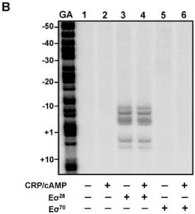
Fig. 3. Sigma factor selectivity and effect of
CRP at the aer promoter in vitro.
A. In vitro transcription from the aer promoter.
The figure shows the transcripts produced in
multi-round in vitro transcription assays using
the aer200 fragment cloned in pSR, incubated
with various concentrations of Es28 or Es70,
and in the presence or absence of 100 nM
CRP and 0.2 mM cAMP, as indicated. The
locations of the s28-dependent aer transcript
and the s70-dependent RNAI control transcript
are indicated.
B. Open complex formation at the aer
promoter. The figure shows the results of
KMnO4 footprinting using an aer200
PstI-HindIII fragment, end-labelled on the
template strand and incubated with a final
concentration of 50 nM Es28 or Es70, in the
presence or absence of 100 nM CRP and
0.2 mM cAMP, as indicated. The gel was
calibrated using a Maxam-Gilbert ‘G + A’
sequencing reaction, and is numbered with
respect to the aer transcript start site.
aer212 and aer211 fragments (Fig. 4, upper three
panels). These fragments were cloned into pRW50, and
expression of the resulting promoter::lacZ fusions was
measured in the CRP+ FliA+, CRP- FliA+ and CRP+ FliA-
backgrounds. The results illustrated in Fig. 4 show that,
when the DNA site for CRP is moved to position -41.5,
aer promoter activity in the CRP+ FliA+ strain is reduced
to a similar level to that observed in the absence of CRP
β-qalactosidase activity
|
CRP* FIiA* |
CRP- FIiA* |
CRP* FIiA- | ||||
|
aer2∞ |
EcoRl crp -35 I_____________J I |
-jθ ∣- |
HMiii |
216 |
12 | |
|
I f 1- I— -4θ.5 |
I |
48 | ||||
|
aer212 |
EooR! |
- Г |
Hindlll |
28 |
50 | |
|
-41.5 |
I |
11 | ||||
|
aer211 |
EooRI _ і j—і і |
Hmdlll |
124 |
35 |
98 | |
|
r LJLJ -61.5 |
I | |||||
|
aer22β |
EcoRI ______ і T |
_ r |
Hindlll |
40 |
60 |
20 |
|
I I I -44.5 |
I | |||||
|
aβr227 |
E∞RI |
»-Γ* |
Hindlll ------------1 |
23 |
4S |
14 |
-54.5
Fig. 4. Effect of moving the DNA site for CRP on activation of the aer promoter. The figure shows schematic diagrams of the wild-type
aer200 promoter fragment, and derivatives in which the DNA site for CRP has been moved to position -41.5 (aer212), -61.5 (aer211), -44.5
(aer226) or -54.5 (aer227). The transcript start sites are indicated by arrows, the locations of the promoter -10 and -35 elements are
indicated by black rectangles and the DNA site for CRP is shaded grey. The figure also shows the b-galactosidase activities (in Miller units)
measured in strain M182 DfliA containing pKXH100 (CRP+ FliA+), strain M182 DfliA Dcrp containing pKXH100 (CRP- FliA+) or strain M182 DfliA
containing ‘empty’ pET21a (CRP+ FliA-), each carrying the different aer promoter::lacZ fusions cloned in pRW50. Cells were grown to late
exponential phase (OD650 0.9-1.1) in LB medium. Data shown are averages from three independent experiments, with a standard deviation of
less than 10%.
© 2009 The Authors
Journal compilation © 2009 Blackwell Publishing Ltd, Molecular Microbiology, 75, 1098-1111
More intriguing information
1. Understanding the (relative) fall and rise of construction wages2. The name is absent
3. Government spending composition, technical change and wage inequality
4. Dynamiques des Entreprises Agroalimentaires (EAA) du Languedoc-Roussillon : évolutions 1998-2003. Programme de recherche PSDR 2001-2006 financé par l'Inra et la Région Languedoc-Roussillon
5. The name is absent
6. Tourism in Rural Areas and Regional Development Planning
7. The name is absent
8. IMPLICATIONS OF CHANGING AID PROGRAMS TO U.S. AGRICULTURE
9. The name is absent
10. Opciones de política económica en el Perú 2011-2015